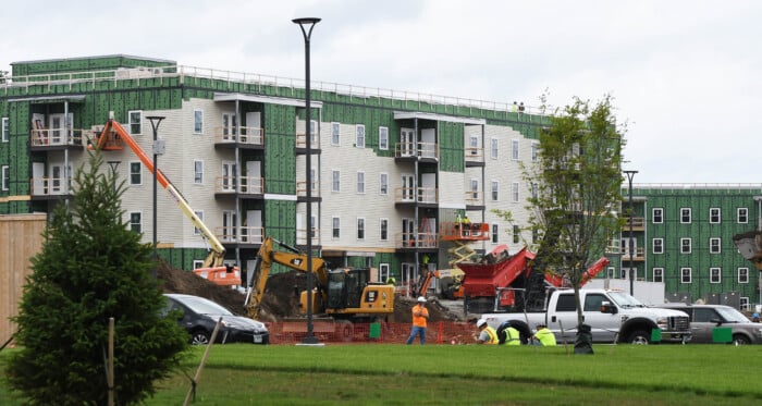
Envision Technologies creates stay, work, play in one Londonderry site
The 110-acre Village at Technology Park off Harvey Road in north Londonderry will include space for industrial, residential, childcare and retail.
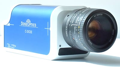
A new imaging system from Hanover-based DoseOptics enables direct imaging of radiation delivery on a routine basis.
The technology works with radiation therapy linacs (linear particle accelerators), capturing both photon and electron beam delivery and providing real-time video rate visualization of the beam shape at the point of incidence and exit. The radiotherapy team can now record and monitor the position and movements while imaging at the site of delivery.
DoseOptics President, Brian Pogue is encouraged about the potential. “We are extremely excited to be able to offer a Cherenkov imaging system to the field of Radiation Oncology where we believe it can change the paradigm of radiation delivery verification, and provide intuitive visualization of the treatment for everyone in the department,” said Dr. Pogue, professor of Engineering Science at the Thayer School at Dartmouth College and professor of Surgery at the Geisel School of Medicine.
Pogue is part of a team of engineers developing Cherenkov imaging technology customized for radiotherapy applications. DoseOptics was aided by a series of grants from the National Cancer Institute at the National Institutes of Health, with the discovery and development occurring at the Thayer School of Engineering at Dartmouth and refined through clinical testing and research at the Norris Cotton Cancer Center at the Dartmouth-Hitchcock Medical Center. The C-Dose system is now available to research customers. Systems have been successfully deployed at leading academic institutions across the United States.
Pogue believes the technology provides a missing piece to today’s therapy. “As new delivery techniques improve and become more and more complex, verification remains a challenge. With C-Dose, medical physicists charged with ensuring delivery accuracy can now literally see what they are doing,” said Pogue in a press release.
Unique time-gating technology ensures that each pulse of the linac contributes to the image recovered, and time-integrating software allows for a cumulative delivered image which is overlaid in real time on the object being irradiated. The camera and software operate remotely to provide an independent check and measurement tool for beam shape and delivery.
“Up until now, we’ve been practicing blind,” says Dr. Lesley Jarvis, MD, PhD, associate professor of Radiation Oncology at Dartmouth Hitchcock Medical Center in Lebanon. “With Cherenkov imaging of phantoms and in clinical trials we have been able to visualize the treatment beams. It’s an intuitive and valuable tool.”
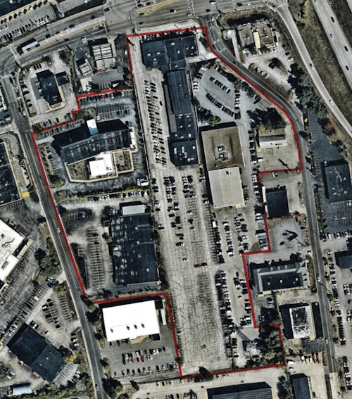
The 110-acre Village at Technology Park off Harvey Road in north Londonderry will include space for industrial, residential, childcare and retail.

At Blueline Advisors in Exeter, chief investment officer Frank Sabin is embracing AI, with the help of the students, to better serve his clients, who have entrusted about $250 million in assets in his care.

As Granite Staters eye ever-increasing purchase prices for a single-family home, state and federal policy makers wrestle with solutions that create more housing supply. Among them is bipartisan federal legislation on housing that is being held hostage by President Donald Trump’s fixation on a bill that would require voters to provide proof of citizenship with such documents as U.S. passports or birth certificates.

Two New Hampshire cities are among the 10 best-run cities in the country, according to a WalletHub analysis of U.S. communities where residents get the most bang for their taxes. Manchester was rated No. 3, while Nashua was close behind at No. 5., based on a “Quality of Services” score that WalletHub developed using 36 metrics across six key service areas — financial stability, education, health, safety, economy and infrastructure/pollution.

Business and event happenings around the state of NH

The Latest is a roundup of the comings and goings of the movers and shakers in NH's business community
A brand new and redesigned Revo Casino and Social House came back to Manchester’s land-use boards this month after the acquisition of additional nearby properties allowed the creation of an expanded vision for the project.
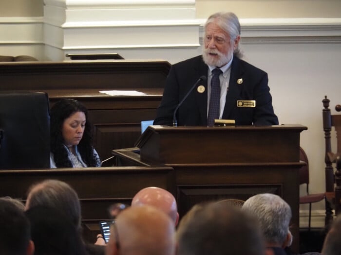
The New Hampshire House and Senate sent three bills to Gov. Kelly Ayotte intended to enable more housing construction, overcoming opposition from the New Hampshire Municipal Association and others.

HEALTH CARE By: DR. STEVEN ANGELO As more Americans live longer, maintaining brain health is becoming an increasingly important part of overall well-being. During Alzheimer’s & Brain Awareness Month, and throughout the year,…